Surgical management of primary hyperparathyroidism: localisation strategies
Primary hyperparathyroidism is common and may result in osteoporosis and renal complications if left untreated. Surgery to remove the hyperfunctioning parathyroid gland(s) is curative. Identifying the responsible gland(s) with imaging before surgery is associated with a higher likelihood of cure and improved patient outcomes. Several imaging modalities are available and selecting the most appropriate one can be challenging for medical practitioners. Three case studies with imaging findings are presented in this article.
- Primary hyperparathyroidism is associated with substantial morbidity but is curable through surgical intervention.
- Minimally invasive parathyroid surgery relies on accurate preoperative imaging studies.
- Neck ultrasound plus four-dimensional CT is now often preferred to [99mTc]sestamibi single-photon emission CT/CT, as it provides greater accuracy and more detailed anatomical assessment.
- [18F]fluorocholine positron emission tomography/CT is a promising novel imaging technique; however, its accessibility is highly limited in Australia at present.
- The choice of preoperative imaging should be guided by patient characteristics, surgeon preference and local availability and expertise.
- Preoperative imaging should be ordered by the treating specialist (an endocrinologist or surgeon) generally only after a decision has been made to proceed with surgical management.
Prevalence and pathogenesis
Primary hyperparathyroidism (PHPT) affects about 1% of the general population and most commonly presents in postmenopausal women.1 It is characterised by elevated serum calcium levels along with elevated or inappropriately normal parathyroid hormone (PTH) levels. In most cases, autonomous production of PTH originates from a single adenoma arising from one of the four parathyroid glands; however, less commonly, there may be multiple adenomas, multigland hyperplasia or, extremely rarely, parathyroid carcinoma.2
Clinical presentation
PHPT can result in substantial morbidity if it is not appropriately diagnosed and managed. Chronic elevation of PTH may lead to symptomatic hypercalcaemia, osteoporosis, renal complications, abdominal symptoms and neuropsychiatric disturbance. The most severe bone complication, osteitis fibrosa cystica, is now almost never seen because of earlier diagnoses.
Diagnosis and further workup
Most cases of PHPT are identified incidentally through biochemical testing of serum calcium and PTH performed for other reasons or in the context of nonspecific symptoms such as fatigue.3 Following the diagnosis, patients with PHPT are typically referred to an endocrinologist or a specialist parathyroid surgeon (endocrine or ear, nose and throat) for assistance with further workup. This involves confirming the diagnosis biochemically, assessing for end-organ complications and deciding between operative or nonoperative management. The diagnosis of PHPT requires repeated measures of serum calcium and PTH, with the exclusion of secondary causes of hyperparathyroidism, including vitamin D deficiency (<75 nmol/L), calcium malabsorption, hypercalciuria (e.g. loop diuretics), renal impairment (estimated glomerular filtration rate <60 mL/min) or medications such as antiresorptives.
The baseline assessment for end-organ complications includes a renal ultrasound, bone density measurement and a thoracolumbar x-ray. A 24-hour urine collection to calculate calcium excretion is key to assess hypercalciuria and exclude familial hypocalciuric hypercalcaemia, as this rare genetic condition can mimic PHPT and does not require treatment. Genetic testing should be considered in all patients under 40 years of age or at any age with additional features, such as a family history of PHPT, in order to exclude conditions such as multiple endocrine neoplasia type 1 or variants in the CDC73 gene (previously known as hyperparathyroidism-jaw tumour syndrome).1 In most cases, only once the decision for surgery has been made should localising imaging be performed to identify the responsible parathyroid gland(s).
Treatment
Surgery is the only curative treatment for PHPT. There is good evidence that successful surgical treatment of PHPT can improve bone health and prevent further decline in renal function; however, the effect on neurocognitive symptoms is less clear.2,4
Identifying which patients require treatment
A proportion of patients with PHPT, particularly those with biochemically mild disease without end-organ complications, require observation only, with monitoring of biochemistry every three to six months. Recent Australian guidelines recommend surgical treatment for the following indications:
- symptoms (Flowchart 1)
- osteoporosis – radiological (low bone density) or clinical (minimal trauma fracture)
- renal complications – nephrolithiasis, nephrocalcinosis, hypercalciuria, deteriorating renal function or a glomerular filtration rate below 60 mL/min in the absence of another explanation
- asymptomatic and under 50 years of age (consider if over 50 years of age with a life expectancy greater than 10 years).1
Further indications for surgical treatment are presented in Flowchart 1.1 In patients who are deemed unfit for surgery, medical therapy with cinacalcet or antiresorptives can be initiated.1 Medical therapy may also be advised for lithium-associated PHPT, given the common occurrence of multigland disease and the high recurrence rates after surgery in this type of PHPT.5

Localising imaging
Localising imaging should not generally influence the decision to offer surgery, but does impact the surgical approach. Identifying the abnormal parathyroid gland(s) on imaging enables targeted surgery – a minimally invasive parathyroidectomy (MIP) – to be performed. MIP requires a small incision of about 2 cm and can be performed as a day case, although overnight admission is usually recommended. In patients with very high surgical risk, the procedure can be performed using local anaesthetic only. Compared with a nontargeted approach, such as bilateral neck exploration, MIP is associated with a shorter duration of operation (about 15 minutes compared with one hour), a reduced length of hospital stay and a lower risk of complications.6 Imaging may also identify ectopic parathyroid glands in the mediastinum, requiring a thoracoscopic approach.
There are several imaging modalities available, and some controversy exists around the exact algorithm by which they should be employed, often influenced by local imaging expertise and preference of the operating surgeon. Because of these nuances, it is recommended that imaging is requested by the treating specialist (an endocrinologist or surgeon). Regardless of the imaging findings, referral to a high-volume specialist parathyroid surgeon is essential to ensure optimal patient outcomes.7
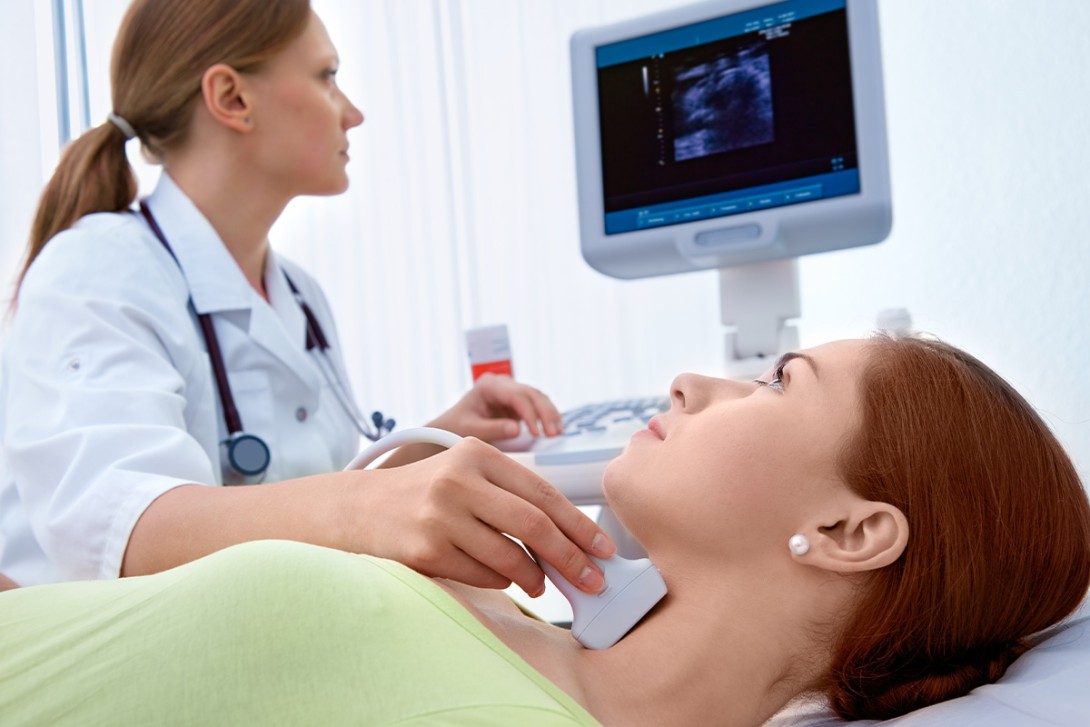
Neck ultrasound
An ultrasound of the neck should be obtained in all cases and is useful in combination with another test to define the relationship of an identified lesion with the thyroid. Ultrasound generally detects only abnormal parathyroid glands; normal parathyroid glands are not typically seen. Neck ultrasounds are quick to perform, cost effective and do not require radiation or intravenous contrast. They may also identify concurrent thyroid pathology that could be addressed at the same time as parathyroid surgery.
However, the limitations of neck ultrasound include reliance on operator skill, inability to identify very small or ectopic parathyroid glands and false negatives because of patient body habitus.8 A fine needle aspirate of an identified lesion to confirm parathyroid tissue is not required and may cause tumour seeding in the rare case of parathyroid carcinoma. The surgeon may perform their own ultrasound before or during the operation.
Sestamibi
In Australia, [99mTc]sestamibi scans usually involve injections of a single radiotracer ([99mTc]sestamibi) with early- and late-phase imaging combined with low-dose single-photon emission CT/CT (SPECT/CT) of the neck and mediastinum, although various protocols exist. Accuracy rates in published direct comparison studies range widely, from 48 to 100%.9 False negative results may be caused by multiglandular disease, adenomas with few oxyphil cells or the presence of a multinodular goitre.
The imaging encompasses a large field of view, enabling the detection of ectopic mediastinal adenomas. Radiation exposure is typically about 7 mSv, equivalent to about three years of background radiation exposure. Sestamibi imaging can take up to four hours to perform, making it time-consuming for the patient. [99mTc] has a short half-life, so it is supplied as the parent compound (99Mo) by the Australian Nuclear Science and Technology Organisation in New South Wales, with generation occurring on site.
Four-dimensional CT
Four-dimensional CT (4DCT) uses multiple-phase contrast-enhanced CT of the neck; the fourth dimension refers to time, with images acquired at different time points. The washout characteristics of the parathyroid glands distinguish them from the background tissue. Higher accuracy rates are reported compared with sestamibi, as well as a shorter imaging time of one hour.9 However, 4DCT exposes patients to greater radiation than sestamibi (typically 12 mSv, or equivalent to about six years of background radiation exposure). The contrast agent can be problematic for patients with allergies or renal impairment. 4DCT is widely available; however, accurate interpretation relies on an experienced radiologist, particularly in combination with feedback from their surgical colleague.
[18F]fluorocholine positron emission technology/CT
[18F]fluorocholine (FCH) positron emission tomography (PET)/CT is a novel imaging technique using PET combined with CT. [18F]FCH was initially used as a radiotracer in prostate cancer imaging; however, it was noted to concentrate in abnormal parathyroid glands. [18F]FCH PET/CT shows very high accuracy rates approaching 95%, is quick to perform (about one hour or less), does not use contrast and exposes patients to less radiation than 4DCT or sestamibi, with the exact dose dependent on the local imaging protocol.9,10 In Europe, [18F]FCH PET/CT has been used widely as a second-line technique where sestamibi, 4DCT, or both, do not localise an abnormal parathyroid gland. In Australia, the availability of [18F]FCH PET/CT was limited to select sites in Perth until mid-2024 when production of [18F]FCH began at QTRaCE, the Radiopharmaceutical Centre of Excellence in the Department of Nuclear Medicine at the Royal Brisbane and Women’s Hospital. Because of the relative stability of [18F]FCH, it has been shipped to other sites in Brisbane and Sydney. Currently, no MBS item number exists for [18F]FCH PET/CT and its use is mainly restricted to externally funded or research settings.
Case studies
Three primary hyperparathyroidism case studies are presented in Box 1, Box 2 and Box 3, with the imaging findings of these cases presented in Figure 1, Figure 2 and Figure 3.


Conclusion
PHPT is an endocrine condition that can be cured with surgery. Preoperative imaging studies can localise the responsible gland(s), facilitating a minimally invasive surgical approach. A neck ultrasound should be obtained in all cases, in conjunction with a second imaging modality. Historically, sestamibi imaging has been the most commonly performed additional investigation; however, 4DCT may now be preferred. [18F]FCH PET/CT is an exciting new modality offering additional benefits, although the availability and cost currently limit use. The exact imaging tests, and at which imaging providers, should be guided by the treating specialist and is often dependent upon local expertise. A suggested approach to the selection of an imaging modality is depicted in Flowchart 2. ET

COMPETING INTERESTS: Dr Wootton and Professor Pattison’s employer, the Royal Brisbane and Women’s Hospital, has received grants or contracts from the Queensland Technology Future Fund and The Royal Australasian College of Physicians.
References
1. Milat F, Ramchand SK, Herath M, et al. Primary hyperparathyroidism in adults-(part I) assessment and medical management: position statement of the Endocrine Society of Australia, the Australian & New Zealand Endocrine Surgeons, and the Australian & New Zealand Bone and Mineral Society. Clin Endocrinol (Oxf) 2024; 100: 3-18.
2. Bilezikian JP. Primary hyperparathyroidism. J Clin Endocrinol Metab 2018; 103: 3993-4004.
3. Walker MD, Silverberg SJ. Primary hyperparathyroidism. Nat Rev Endocrinol 2018; 14: 115-125.
4. Rubin MR, Bilezikian JP, McMahon DJ, et al. The natural history of primary hyperparathyroidism with or without parathyroid surgery after 15 years. J Clin Endocrinol Metab 2008; 93: 3462-3470.
5. Mifsud S, Cilia K, Mifsud EL, Gruppetta M. Lithium-associated hyperparathyroidism. Br J Hosp Med (Lond) 2020; 81: 1-9.
6. Jinih M, O’Connell E, O’Leary DP, Liew A, Redmond HP. Focused versus bilateral parathyroid exploration for primary hyperparathyroidism: a systematic review and meta-analysis. Ann Surg Oncol 2017; 24: 1924-1934.
7. Chander NR, Chidambaram S, Van Den Heede K, DiMarco AN, Tolley NS, Palazzo FF. Correlation of preoperative imaging findings and parathyroidectomy outcomes support NICE 2019 guidance. J Clin Endocrinol Metab 2022; 107: e1242-e1248.
8. Miller JA, Gundara J, Harper S, et al. Primary hyperparathyroidism in adults-(part II) surgical management and postoperative follow-up: position statement of the Endocrine Society of Australia, The Australian & New Zealand Endocrine Surgeons, and The Australian & New Zealand Bone and Mineral Society. Clin Endocrinol (Oxf) 2024; 101: 516-530. Epub 2021.
9. Lee SW, Shim SR, Jeong SY, Kim SJ. Direct comparison of preoperative imaging modalities for localization of primary hyperparathyroidism: a systematic review and network meta-analysis. JAMA Otolaryngology Head Neck Surg 2021; 147: 692-706.
10. Quak E, Cavarec M, Ciappuccini R, Girault G, Rouzier R, Lequesne J. Detection, resection and cure: a systematic review and meta-analysis of 18F-choline PET in primary hyperparathyroidism. Q J Nucl Med Mol Imaging 2023; 67: 122-129.
Single article purchases are temporarily unavailable due to site maintenance.
If you would like to purchase an article during this time, please email us at [email protected] with the article details and we'll assist you directly. We'll also let you know when online purchasing is available again.
Thank you for your patience and understanding.