Endocrine causes of osteoporosis. Part 3: Premature ovarian insufficiency
Osteoporosis
Menopause
Endocrine causes of osteoporosis, such as primary hyperparathyroidism, hypogonadism, glucocorticoid excess, acromegaly, Cushing’s syndrome and hyperthyroidism, are well-recognised risk factors for decreased bone mineral density. Optimal management of bone health in people with these conditions involves treating the underlying hormonal disease and assessing the need for specific bone preservation therapy. This three-part series discusses three cases to highlight the detrimental impact of specific endocrine diseases on bone health.
- Premature ovarian insufficiency (POI) is defined as amenorrhoea or oligomenorrhoea before the age of 40 years associated with a follicle stimulating hormone level of more than 40 IU/L in women who previously had normal menstrual cycles.
- Menopausal hormone therapy is recommended in women with POI for the treatment and maintenance of bone health, unless contraindicated.
- Optimal management of lifestyle factors, such as smoking cessation, adequate dietary calcium intake and exercise, is also recommended.
- Women with POI and suboptimal bone health despite use of appropriate menopausal hormone therapy warrant referral to an endocrinologist for consideration of nonhormonal antiresorptive therapy.
Premature ovarian insufficiency
Case scenario
Ms Robinson, a 30-year-old woman, was referred to an endocrinologist by her GP for assessment of secondary amenorrhoea. She went through menarche at 16 years of age and started on the oral contraceptive pill in her late teens for management of oligomenorrhoea and menorrhagia. At 26 years of age, she stopped the oral contraceptive pill as she and her partner wished to have a family. Although light menses recurred eight months later, her menses were initially erratic and then ceased. She subsequently developed troublesome hot flushes.
Ms Robinson had no family history of premature menopause or autoimmune disease and no personal history of prior infections or other comorbidities. She was a nonsmoker and only consumed alcohol intermittently. Examination revealed Tanner stage V breast and pubic hair development. Her body mass index was 22 kg/m2.
She was investigated for causes of secondary amenorrhoea. Her follicle stimulating hormone (FSH) level was found to be elevated at 64 IU/L (normal 1.8–2.3 IU/L for premenopausal women) and her oestradiol level was 44 pmol/L (normal 100–1570 pmol/L in premenopausal women). These results were confirmed on repeat testing several weeks later. A pelvic ultrasound revealed small ovaries with absence of ovarian follicles and her endometrial thickness was 2 mm. Karyotyping and fragile-X premutation test results were normal. Adrenal and thyroid antibodies were not detected. Ms Robinson was diagnosed with idiopathic premature ovarian insufficiency (POI).
Dual-energy x-ray absorptiometry (DXA) showed a Z-score of –2.3 at the lumbar spine and –1.8 at the right total hip, consistent with reduced bone mineral density (BMD). She was commenced on estradiol 100 mcg/24 hours transdermal patch twice a week and norethisterone 2.5 mg orally on days 1 to 12 of the menstrual cycle. She was also referred to a reproductive specialist and psychologist. DXA was repeated after two years and showed the right total hip and lumbar spine scores had improved by 4.5% and 5%, respectively.
Discussion
The global prevalence of POI is 1 to 3.7%.1,2 The International Menopause Society defines POI as amenorrhoea or oligomenorrhoea (less than eight menstrual cycles per year) before the age of 40 years associated with an FSH level of more than 40 IU/L in women who previously had normal menstrual cycles. POI may occur due to iatrogenic causes such as bilateral oophorectomy, prior chemotherapy and abdominopelvic radiotherapy. Causes of spontaneous POI include genetic abnormalities, such as Turner syndrome and Fragile-X syndrome, rare enzyme deficiencies, infections such as mumps oophoritis and other associated infections including HIV, tuberculosis, cytomegalovirus and herpes zoster virus. Of women with POI, 10 to 27% have associated autoimmune disease (adrenal autoimmunity, coeliac disease and thyroid autoimmunity are not uncommon).3 Although women may present with vasomotor symptoms, menstrual disturbances or infertility, maintaining bone health remains an important objective.2
There is a complex interplay between normal sex steroid levels and the musculoskeletal system that leads to increased bone mass, improved bone microarchitecture and improved muscle strength, all of which contribute to reduced fracture risk.4,5 In the presence of oestrogen deficiency, rapid decline in BMD occurs of the order of 2.4% per year in the first five years with persistent losses at a lower rate up to 10 years after menopause.6
Prevention and management of osteoporosis in women with POI can be challenging due to the scarcity of evidence in this cohort. Much of the evidence is based on extrapolation of data from studies performed in postmenopausal women. The association between oestrogen deficiency and reduced BMD is well established, but larger studies are required to establish fracture risk in women with POI.
The optimal management of lifestyle factors such as smoking cessation, adequate dietary calcium intake and exercise is recommended (Box 1). Weight-bearing exercise has demonstrated efficacy in improving femoral neck BMD in men and postmenopausal women, whereas a combination of strength and aerobic exercise may be beneficial for improvement of lumbar spine BMD.7,8
In the absence of contraindications, menopausal hormone therapy (MHT) until the natural age of menopause (about 51 years) is recommended to reduce the detrimental effects of oestrogen loss on bone. This recommendation by the European Society of Human Reproduction and Embryology (ESHRE) is based on smaller prospective studies that demonstrate stable or improved BMD in women receiving MHT.9 The benefits of MHT are not just limited to bone health. Women taking MHT may experience reduced frequency of vasomotor symptoms, improved sexual health with reduced vaginal dryness and improved libido. MHT also improves the cardiovascular risk profile in women with POI through beneficial effects on lipid profile and insulin resistance.2 The ESHRE guidelines provide a comprehensive overview of the management of women with POI.9 A list of available MHT preparations can be found on the Australian Menopause Society website.10
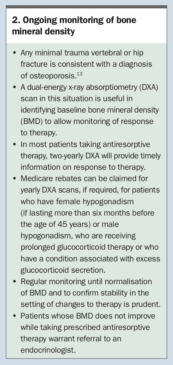
Delay in estrogen replacement therapy is associated with higher rates of bone loss in women with POI.11 In women not wanting to conceive, contraception is recommended due to the small proportion of women (5%) with POI who are able to conceive in the setting of physiological oestrogen levels.12 There are limited data to guide BMD monitoring in women with POI after commencement of MHT. Biennial measurement of BMD using DXA, for monitoring of response to therapy, is reasonable (see Box 2 for information on ongoing monitoring of BMD, which applies to all patients with osteoporosis).13
In the event of persistently low BMD, loss of BMD or new fractures despite use of MHT, specialist review should be considered. ET